

Ph.D. em
neurologia infantil
Médica e pesquisadora, atuando no atendimento de crianças e adolescentes com Epilepsia, Transtorno do Espectro do Autismo (TEA), Transtorno do Déficit de Atenção e Hiperatividade (TDAH), Transtorno Opositor Desafiante (TOD), Transtorno do Desenvolvimento Intelectual (DI), Transtorno do Processamento Sensorial (TPS), Transtorno do Desenvolvimento da Linguagem (TDL), Transtornos de Aprendizagem, Síndromes Raras, Altas Habilidades e Superdotação (AHSD), entre outros. Também atua no acolhimento de adultos em busca do diagnóstico atípico tardio.
É membro do Serviço de Neurologia Infantil do Hospital Moinhos de Vento, em Porto Alegre – RS, e o Hospital Albert Einstein, em São Paulo -SP. Atualmente, atua em consultório particular, organizando plano terapêutico multidisciplinar individualizado para cada paciente, com supervisão médica e reavaliação continuada. É membro do INSCER, Instituto do Cérebro da PUCRS – Pontifícia Universidade Católica do Rio Grande do Sul.
Consultora credenciada do PlayProject no Brasil, é responsável pelo curso Treinamento Iplay, um programa destinado a pais, professores, cuidadores, familiares, educadores, terapeutas, a quem interessar, em intervir precocemente na reabilitação de crianças autistas, através do brincar.
Idealizadora do Projeto Consulta Social, uma iniciativa de acolher famílias de baixa renda, disponibilizando atendimento médico gratuito as pessoas acometidas pelos Transtornos do Neurodesenvolvimento.
PLAY Project™ — PORTO ALEGRE, BRASIL
Consultora credenciada internacional
O PLAY Project ™️ é um programa intensivo de intervenção precoce baseado em evidências, para crianças com Transtorno do Espectro do Autismo, implementado para pais.
CONHEÇA O PROJETO2020 — até o momento

INSCER — Instituto do Cérebro — Porto Alegre, Brasil.
Pesquisadora
Desenvolvendo um estudo sobre recém nascidos de alto risco.
CONHEÇA O INSTITUTO2018 — até o momento

Hospital Moinhos de Vento — Porto Alegre, Brasil.
Membro do serviço de Neurologia
CONHEÇA A INSTITUIÇÃO2010 — até o momento

Formação.
Médica, mestre, doutora e pós-doutura em Neurociencias na Pontifícia Universidade Católica do Rio Grande do Sul e pela UCLA – School of Medicine (EUA), com Residência Médica em Pediatria na UFCSPA, e especialista em Neurologia Infantil pela Fundação Oswaldo Cruz. Atuamente aborda a temática “Recém nascidos de alto risco” como pesquisadora do INSCER – Instituto do Cérebro da PUCRS. Ao longo dos anos, esteve continuamente desenvolvendo conteúdo científico que é compartilhado no campo médico/acadêmico através de artigos, projetos e publicações.
PUCRS — Pontíficia Universidade Católica do Rio Grande do Sul
Pós-Doutorado
2011 — 2014
Bolsista do(a): Coordenação de Aperfeiçoamento de Pessoal de Nível Superior, CAPES, Brasil.
Grande Área: Ciências da Saúde
Área: Medicina
Subárea: neurologia
PUCRS — Pontíficia Universidade Católica do Rio Grande do Sul
Doutorado em Medicina e Ciências da Saúde
2008 — 2010
Com período sanduíche em UCLA School of Medicine (Orientador: Gary W Mathern).
Título: Relação entre Injúria Precipitante Inicial e Dano Hipocampal na Displasia Cortical Focal tipo I
Ano de obtenção: 2010
Orientador: Magda Lahorgue Nunes
Bolsista do(a): Coordenação de Aperfeiçoamento de Pessoal de Nível Superior, CAPES, Brasil.
Palavraschave: epilepsy; focal cortical dysplasia; Seizure; surgery; initial precipitating injury; dual pathology.
Grande Área: Ciências da Saúde
Área: Medicina
Subárea: Neurologia
Setores de atividade: Saúde humana e serviços sociais
PUCRS — Pontíficia Universidade Católica do Rio Grande do Sul
Mestrado em Medicina e Ciências da Saúde
2006 — 2007
Título: Efeitos da desnutrição precoce e de crises convulsivas na memória espacial do rato imaturo
Ano de Obtenção: 2007.
Orientador: Magda Lahorgue Nunes
Bolsista do(a): Coordenação de Aperfeiçoamento de Pessoal de Nível Superior, CAPES, Brasil.
Palavraschave: desnutrição; epilepsia.
Grande área: Ciências da Saúde
Setores de atividade: Saúde Humana
FIOCRUZ — Fundação Oswaldo Cruz
Especialização em Neurologia Infantil
2004 — 2006
Bolsista do(a): Fundação Oswaldo Cruz, FIOCRUZ, Brasil.
UFCSPA — Fundação Universidade Federal de Ciências da Saúde de Porto Alegre
Especialização Residência médica
2002 — 2003
Residência médica em: Pediatria
Número do registro: 296CNRM.
Bolsista do(a): Ministério da Educação, MEC, Brasil.
PUCRS — Pontíficia Universidade Católica do Rio Grande do Sul
Graduação em Medicina
1996 — 2001
Título: HIV e Demência.
Orientador: Alfredo Cataldo Neto
NUHS — North Union High School
Formação Complementar
1993 — 1993
Curso de Nível Secundário.
Projetos, estudos,
artigos e mais.
Confira as pesquisas que já levaram a principais publicações na revista Neurology (EUA), inclusive sendo capa da revista, e no International Journal of Developmental Neuroscience.

Assessment and surgical outcomes for mild type I and severe type II cortical dysplasia: A critical review and the UCLA experience
Recent findings on the clinical, electroencephalography (EEG), neuroimaging, and surgical outcomes are reviewed comparing patients with Palmini type I (mild) and type II (severe) cortical dysplasia. Resources include peer-reviewed studies on surgically treated patients and a subanalysis of the 2004 International League Against Epilepsy (ILAE) Survey of Pediatric Epilepsy Surgery. These sources were supplemented with data from University of California, Los Angeles (UCLA). Cortical dysplasia is the most frequent histopathologic substrate in children, and the second most common etiology in adult epilepsy surgery patients. Cortical dysplasia patients present with seizures at an earlier age than other surgically treated etiologies, and 33-50% have nonlocalized scalp EEG and normal magnetic resonance imaging (MRI) scans. 2-((18)F)Fluoro-2-deoxy-D-glucose positron emission tomography (FDG-PET) is positive in 75-90% of cases. After complete resection, 80% of patients are seizure free compared with 20% with incomplete resections. Compared with type I, patients with type II cortical dysplasia present at younger ages, have higher seizure frequencies, and are extratemporal. Type I dysplasia is found more often in adult patients in the temporal lobe and is often MRI negative. These findings identify characteristics of patients with mild and severe cortical dysplasia that define surgically treated epilepsy syndromes. The authors discuss future challenges to identifying and treating medically refractory epilepsy patients with cortical dysplasia.
Clique aqui para acessar o conteúdo
Effects of early malnutrition, isolation and seizures on memory and spatial learning in the developing rat
Purpose: In this study we evaluated the effects of undernourishment and seizures on memory and spatial learning in a model of developing brain.
Experimental procedures: Male Wistar rat pups were allocated to one of six experimental groups: nourished control (NC), nourished recurrent seizures (NRS), nourished status epilepticus (NSE), undernourished control (UC), undernourished recurrent seizures (URS) or undernourished status epilepticus (USE). The UC, URS and USE groups were maintained on a starvation regimen from postnatal day 2 (P2) to postnatal day 15 (P15). URS and NRS groups suffered three daily Flurothyl-induced seizures from P2 to P4. The USE and NSE groups suffered a status epilepticus (SE) on P15. Beginning on P21 all groups were trained in the Morris water maze. At P30 the animals were sacrificed and their brains weighed.
Results: Our data indicate that early undernourishment does not alter seizure susceptibility at P15, but diminishes body and brain weight (p < 0.001), whereas seizures diminish body (p < 0.001) but not brain weight (p = 0.972). In the Morris water probe test we have observed that undernourished rats spent less time in the target quadrant than nourished animals (p < 0.001). Also, rats submitted to recurrent seizures and rats submitted to status epilepticus spent less time in the target quadrant than seizure-free animals (p = 0.001). There was a significant interaction between undernourishment and seizure (p = 0.013).
Discussion: Our findings show that undernourishment and seizures have an additive detrimental effect on body and brain weight as well as on spatial memory.
Clique aqui para acessar o conteúdo
Clinical and electroencephalographic characteristics of benign occipital epilepsy of childhood in two tertiary Brazilian hospitals
This study intended to investigate the clinical and electroencephalographic benign occipital epilepsy of childhood (BOEC) characteristics in a population sample of patients from two tertiary Brazilian hospitals. We analyzed retrospectively 4912 electroencephalograms (EEGs) records, and the included patients were submitted to a new clinical and EEG evaluation. Were included 12 (0.92%) patients; 4 (33.3%) with criteria for early BOEC; 6 (50%) for late form and 2 (16.7%) with superimposed early and late onset forms. After new investigation, 2 (16.7%) had normal EEG; 4 (33.3%) had paroxysms over the occipital region; 3 (25%) over the temporal posterior regions and 3 (25%) over the posterior regions. Sharp waves were the predominant change, occurring in 8 (66.6%); spike and slow wave complexes in 1 (8.3%) and sharp and slow wave complexes in 1 (8.3%). Vomiting, headache and visual hallucinations were the most common ictal manifestations, presented in 100% of patients with superimposed forms. Vomiting were absent in the late form and headache was present in all forms of BOEC.
Bilateral Rasmussen’s encephalitis associated with type II focal cortical dysplasia: Dormant ‘second’ epileptogenic zone in contralateral disease
Rasmussen’s encephalitis (RE) is an inflammatory, probably autoimmune disorder manifested by refractory seizures and progressive deterioration of one cerebral hemisphere [1].
Here, we describe the unfortunate history of a girl with a progressive disorder which, upon clinical, neuroimaging, and histopathological evaluation, proved to be bilateral RE associated with type II focal cortical dysplasia. Whether the second pathology is relevant for the extent of the disease is discussed.
We demonstrated histopathological evidence of RE and type II FCD in the left hemisphere, which led to EPC on the right hemibody at presentation. In addition, there was unequivocal progressive cortical and subcortical atrophy of the right hemisphere, which accounted for the EPC on the left hemibody. This is highly compatible with RE (+/− FCD) in the right hemisphere as well. Although the association of FCD and RE – as well as the occasional occurrence of bilateral RE – has already been reported [3], [4], [5], this is the first such case in which bilateral RE and FCD co-occur.
Clique aqui para acessar o conteúdo
An 18-year follow-up of seizure outcome after surgery for temporal lobe epilepsy and hippocampal sclerosis
Objectives To evaluate the very long-term clinical outcome of surgery for mesial temporal lobe epilepsy and unilateral hippocampal sclerosis (MTLE/HS) without atypical features. The impact of surgical technique and postoperative reduction of medication on this outcome was investigated.
Design Prospective longitudinal cohort follow-up study for up to18 years.
Setting Epilepsy surgery centre in a university hospital.
Patients 108 patients who underwent unilateral MTLE/HS.
Intervention Surgery for MTLE/HS.
Main outcome measure Engel classification (I). Clinical evaluations were based on systematic interviews in person or by phone. Kaplan-Maier survival curves estimated the probability of remaining seizure free. The impact of medication management in the postoperative outcome was analysed using Cox regression.
Results The probability of remaining completely seizure-free at 12 and 18 years after MTLE/HS surgery was 65% and 62%, respectively. The risk of having any recurrence was 22% during the first 24 months and increased 1.4% per year afterwards. Type of surgical technique (selective amygdalohippocampectomy vs anterior temporal lobectomy) did not impact on outcome. Remaining on antiepileptic drugs and history of generalised clonic seizure diminished the probability of remaining seizure free.
Conclusions MTLE/HS surgery is able to keep patients seizure free for almost up to two decades. Removal of the neocortex besides the mesial portion of the temporal lobe does not lead to better chances of seizure control. These findings are applicable to the typical unilateral MTLE/HS syndrome and cannot be generalised for all types of TLE. Future longitudinal randomised controlled studies are needed to replicate these findings.
Clique aqui para acessar o conteúdoEffects of undernourishment, recurrent seizures and enriched environment during early life in hippocampal morphology
It has been recently shown that enriched environment led to a significant benefit in learning and retention of visual–spatial memory, being able to reverse the cognitive impairment generated by undernourishment and recurrent seizures. We investigated the hippocampal morphological effects of recurrent seizures and undernourishment early in life in Wistar rats and the possible benefits produced by the enriched environment in these conditions. The morphological parameters stereologically evaluated were hippocampal volume, thickness of pyramidal stratum of the CA1 subfield and neuronal and glial densities in the same subfield. Male Wistar rats were divided into eight groups including nourished, nourished + enriched environment, nourished + recurrent seizures, nourished + recurrent seizures + enriched environment, undernourished, undernourished + enriched environment, undernourished + recurrent seizures and undernourished + recurrent seizures + enriched environment. Undernourishment model consisted in nutritional deprivation regimen from post-natal day 2 (P2) to P15. From P8 to P10, recurrent seizures group were induced by flurothyl three times per day. Enriched environment groups were exposed between P21 and P51. Our main findings were: (1) animals submitted to the enriched environment showed an increased hippocampal volume; (2) enriched environment promotes increases in the thickness of the pyramidal layer in hippocampal CA1 subfield in animals nourished and undernourished with recurrent seizures; (3) undernourishment during early development decreased neuronal density in CA1 and CA3 subfields. Our findings show that these three conditions induces important changes in hippocampal morphology, the most deleterious changes are induced by undernourishment and recurrent seizures, while more beneficial morphological changes are produced by enriched environment.
Clique aqui para acessar o conteúdo
“Mirror EPC” Epilepsia partialis continua shifting sides after rolandic resection in dysplasia
Background: Epilepsia partialis continua (EPC) is a life-threatening condition often caused by focal cortical dysplasia (FCD). Resection of the motor cortex is contemplated in the hope that the trade-off between a severe motor deficit and complete seizure control justifies the procedure.
Methods: Report of 3 patients with EPC due to histologically confirmed FCD, who underwent resection of the motor cortex under acute electrocorticography.
Results: All had re-emergence of medically intractable EPC in the other side of the body after rolandic resection. Two patients died and the third continues with refractory attacks.
Conclusion: In some instances, EPC due to FCD may shift sides and re-emerge in the contralateral, previously asymptomatic, hemibody. A mechanism of disinhibition by surgery of a suppressed contralateral and homologous epileptogenic zone is speculated.
Clique aqui para acessar o conteúdo
Dysplastic Cerebellar Epilepsy: Complete Seizure Control Following Resection of a Ganglioglioma
Subcortical epilepsy has been a controversial issue, partially settled by evidence showing seizure generation in hypothalamic hamartomas and also by reports of seizures caused by cerebellar lesions. We report 4-year-old girl with right hemifacial seizures and autonomic phenomena, in whom MRI showed an irregular mass in the right cerebellar peduncle. Despite several unremarkable video-EEG recordings, seizure origin in the lesion was hypothesized. Complete resection was feasible, histopathology showed a ganglioglioma, and she has been seizure free for 3 years. A fine line separates these developmental tumors from focal cortical dysplasia, and the homogeneous presentation of this entity led us to propose the terminology dysplastic cerebellar epilepsy.
Clique aqui para acessar o conteúdoSelective posterior callosotomy for drop attacks. A new approach sparing prefrontal connectivity
Objective: To evaluate a novel approach to control epileptic drop attacks through a selective posterior callosotomy, sparing all prefrontal interconnectivity.
Methods: Thirty-six patients with refractory drop attacks had selective posterior callosotomy and prospective follow-up for >4 years. Falls, episodes of aggressive behavior, and IQ were quantified. Autonomy in activities of daily living, axial tonus, and speech generated a functional score ranging from 0 to 13. Subjective effect on patient well-being and caregiver burden was also assessed.
Results: Median monthly frequency of drop attacks decreased from 150 to 0.5. Thirty patients (83%) achieved either complete or >90% control of the falls. Need for constant supervision decreased from 90% to 36% of patients. All had estimated IQ below 85. Median functional score increased from 7 to 10 (p = 0.03). No patient had decrease in speech fluency or hemiparesis. Caregivers rated the effect of the procedure as excellent in 40% and as having greatly improved functioning in another 50%. Clinical, EEG, imaging, and cognitive variables did not correlate with outcome.
Conclusions: This cohort study with objective outcome assessment suggests that selective posterior callosotomy is safe and effective to control drop attacks, with functional and behavioral gains in patients with intellectual disability. Results are comparable to historical series of total callosotomy and suggest that anterior callosal fibers may be spared.
Classification of evidence: This study provides Class III evidence that selective posterior callosotomy reduces falls in patients with epileptic drop attacks.
Clique aqui para acessar o conteúdo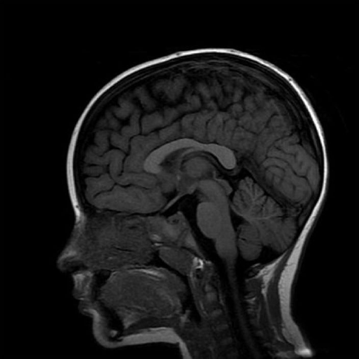
Thermoregulatory Instability in Childhood: Linking the Normal Brain to Hypothalamic Storm
Central core temperature is tightly controlled by hypothalamic centers, a feature that makes sudden changes in body temperature very unusual. A dysfunction of these hypothalamic pathways leads to Shapiro’s syndrome, comprising spontaneous hypothermia, hyperhidrosis, and corpus callosum dysgenesis. Although it may affect any age, usually it presents in childhood. Variants to this syndrome with completely normal brain anatomy have been consistently reported, expanding the clinical spectrum of the syndrome. Herein, we report the case of a 4-year-old girl with Shapiro’s syndrome and unaffected corpus callosum.
Clique aqui para acessar o conteúdoVamos conversar?
Envie um e-mail para martahemb@gmail.com
Para agendamentos, utilize os telefones abaixo:
Consultório
(51) 99655-7793
Rua Miguel Tostes, 201 / Sala 602
Rio Branco, CEP 90430-061
Porto Alegre — RS, Brasil
Assessoria de Comunicação
Para agendar Entrevistas, Palestras e Lives:
Rochele Zago – (51) 98168-5096
Instagram: @rochelezago